
Placement of PD Catheters
Prior to placement of a PD catheter a complete medical and surgical history and physical exam should take place. Situations that may complicate catheter placement include hernias, abdominal mesh, organomegaly, previous kidney transplantation, and/or previous abdominal surgeries. If time allows, a bowel preparation and pre-operative fasting is recommended. In preparation for surgery the patient should shower with disinfectant soap and prophylactic antibiotics may be administered.
The best placement of the catheter within the abdominal wall was thoroughly studied by Tenckhoff, who included himself among the study subjects (1). The dacron cuffs are placed within the rectus muscle and in the subcutaneous tissue of the anterior abdominal wall. The cuff induces a classic inflammatory reaction characterized by formation of fibrin clots, ingrowth of granulocytes and fibroblasts and granulomata with giant cells. The resulting fibrous plug represents a curious case of beneficial bioincompatibility. It prevents bacteria from entering the subcutaneous space from the skin surface into the peritoneum. At the skin exit site, stratified squamous epithelium grows along the surface of the catheter ending in granulation tissue near the pre-peritoneal cuff. At the peritoneal surface, simple squamous epithelium grows around the catheter, penetrating the abdominal wall and ending at the deep cuff, resulting in a smooth surface surrounding the catheter (2).
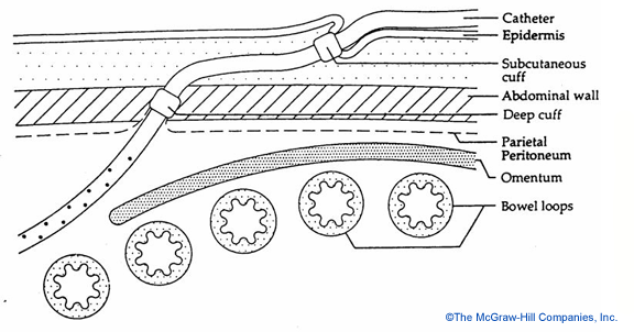
Optimal Catheter Placement
Used with permission from reference 2.
The midline penetration through the linea alba was commonly used in the past due to fewer vascular structures; however, it is now mostly reserved for acute catheters and as a last resource only. The preferred primary penetration sites for chronic catheters are the lateral or paramedian insertions through the rectus muscle since they are associated with better outcomes and a lower rate of complications (3-5).
The exit-site placement is critical for good outcomes. An arcuate subcutaneous tunnel and a downward exit site for the catheter was first proposed by Tenckhoff in 1968 to maintain the internal and external portions of the catheter in a caudad direction and to foster drainage from the subcutaneous tunnel and prevent infection (1). The advantages of this practice in reducing the number and severity of infections have been reported by several authors (6-9). The exit site should precisely fit the catheter diameter in order to avoid extrusion of the external cuff if it is too large, or pressure necrosis if too tight. This can be easily accomplished using a commercially available perforator or by a single pass with a Parker blade # 11. The subcutaneous cuff should be 1-2 cm from the skin after healing.
Methods of Implantation
Catheter insertion techniques may be classified as surgical (dissective) or percutaneous. Percutaneous methods may be further categorized according to whether the procedure is performed by blind or guided techniques. Blind insertions are characterized by penetration of the abdominal cavity without visual assistance. Blind catheter implantation may be performed using the Trocar Method or the Modified Seldinger Technique (10). In contrast, guided methods allow for visualization of the peritoneal cavity prior to catheter placement and may incorporate the use of fluoroscopy, peritoneoscopy or laparoscopy. Each method is associated with unique advantages and limitations that should be considered during the selection process. In addition, operator proficiency greatly contributes to the overall success of catheter insertion. Therefore, decisions regarding technique selection should also be based on the expertise of the surgeon.
Surgical Placement
Traditional surgical implantation is the most common method of catheter placement. This method involves direct incision of the skin, rectus muscle and parietal peritoneum, followed by insertion of the catheter and stylet (11). The procedure typically requires light general anesthesia. The principal advantages are precise placement of the catheter tip, optional fixation and lysis of adhesions and omentectomy. Despite these benefits, surgical placement techniques increase the risk for pericatheter leaks when compared to percutaneous implantation methods.
Blind Trocar Method
The earliest clinically successful percutaneous catheter placement incorporated the use of a large bore trocar developed by Tenckhoff. A trocar is an instrument that is used to penetrate the abdominal cavity during catheter insertion. Blind trocar insertion is a relatively simple technique that is usually performed with local anesthesia. Major advantages of this technique are the option for rapid catheter insertion and the ability to perform the procedure at the patient’s bedside.
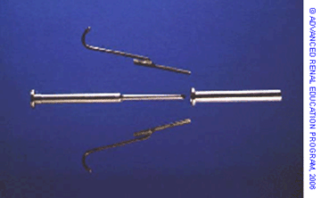
Tenckhoff Trochar
Courtesy of Dr. J.A. Diaz-Buxo.
Disadvantages include a large penetration site diameter and a lack of visualization of the peritoneum to guide insertion. As a result, blind techniques are accompanied by an increased risk of bowel perforation especially in obese patients and in those with a history of abdominal surgeries (10).
Modified Seldinger Technique
Percutaneous implantation has been further simplified with a modified Seldinger technique similar to that used for insertion of vascular catheters (12). Although this is a blind procedure, the risk of perforation is relatively low since it uses a simple needle for penetration followed by dilatation with a blunt plastic dilator. The technique has similar risks and benefits as the Blind Trocar Method; however, the location of the catheter can be improved with the use of imaging assistance (e.g. fluoroscopy).
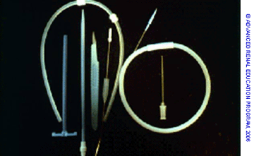
Seldinger Technique Disposables
Courtesy of Dr. J.A. Diaz-Buxo.
Fluoroscopy-Assisted Placement
In contrast to the Blind and Modified Seldinger techniques, fluoroscopy-guided insertion allows for visualization of the peritoneum during catheter placement. This technique incorporates the use of contrast media that is inserted into the peritoneum to allow for identification of peritoneal structures. Fluoroscopy-guided catheter insertion allows for confirmation of the location of the catheter within the peritoneum. The technique is limited by the poor quality of the generated image, preventing identification of adhesions or omentum that may impede catheter function.
Peritoneoscopic Technique
Peritoneoscopic techniques involve the insertion of a peritoneoscope, an instrument that is used to view the peritoneal cavity prior to catheter insertion. The procedure may be performed under local anesthesia and may allow for early identification of imposing structures and impediments to proper catheter function.
Laparoscopic/mini-laparoscopic Technique
Laparoscopic implantation with or without omentopexy (13)is a minimally invasive approach that is usually associated with less pain and quicker return to full activities, especially in obese patients. It offers complete visualization of the catheter insertion process and permits selective proactive intervention for catheter migration, omental entrapment and obstructive adhesions. It also allows for the diagnosis and treatment of previously unsuspected herniae. Limitations of laparoscopic procedures include a requirement for general anesthesia (not appropriate for all patients) and the delivery of two puncture wounds to the peritoneum (for the scope and catheter) (10).
Alternative Placement Techniques
Moncrief-Popovich Technique
Subcutaneous burial of the external segment of the catheter to prevent colonization of the catheter by skin bacteria and promote attachment of the cuff to the tissue prior to exteriorization has been described and recently tested by several experienced clinicians with encouraging results (14). The initial reports by the developers claimed a reduction in the rate of peritonitis and colonization of bacterial biofilms in the catheter segments between the two cuffs (15). However, a controlled randomized study failed to confirm these claims (16). A possible reason for the failure to reduce the incidence of infectious complications may be the inability of the body to provide an effective “seal” around the external cuff. Therefore, upon exteriorization of the catheter, the process of healing starts all over again. Prischl et al. have also reported a high incidence of seromas, subcutaneous hematomas and fibrin thrombi postoperatively with this technique (17).
Extended Dialysis Catheters
Extended dialysis catheters have been developed to allow placement of the exit site in remote places and preferably in the presternal area (18). This is particularly useful for obese patients, the rare patient with a stoma and those with other sources of potential contamination in the anterior abdominal wall. The commercially available kits contain catheters of various configurations, a presternal extension tube and a titanium connector to join the aforementioned parts.
The main advantages of these catheters are better wound healing and immobilization, decreased pressure by garments, increased distance from ostomy sites, easier exit site care, lower frequency of exit site infections and peritonitis in selected cases. The main disadvantage is the possibility of disconnection inside the subcutaneous tunnel. Body image is thought to be improved by some patients but definitely worse by others.
A six-year non-randomized prospective study comparing swan-neck presternal catheters with swan-neck abdominal catheters showed good tolerance of the presternal catheters but no significant differences in survival or peritonitis rates (19). Similarly, a retrospective comparison by a single surgeon comparing a standard, double-cuff Tenckhoff catheter (n=46) or a swan-neck presternal catheter (n=14) reported a lower, but non-significant difference in the rate of exit-site infections (20).
Self-Locating or Front Loading Catheters
Several European reports describing a PD catheter with either a 12-g tungsten cylinder embedded in the silicone or stainless steel weights attached to the distal end of the catheter suggest better catheter survival and less migration as compared to standard catheters (21-24). Although the data from clinical trials are limited, the results are consistently positive. Further evaluation of this simple modification to the peritoneal catheter is warranted.
Proper catheter insertion is critical to the initial success of PD therapy. Poor placement technique may lead to a variety of complications such as outflow obstruction, pericatheter leakage, infection, and bowel perforation and may limit the long-term viability of PD as a therapy option for patients. Therefore, it is essential that catheter insertion be performed by an experienced clinician who has carefully considered individual patient characteristics that may influence the selection of one implantation method over another.
Table 1: Complications associated with different techniques25-35
|
Method |
Author (Year) |
Catheters (n) |
Leak (%) |
Flow dysfunction (%) |
Leak plus flow dysfunction (%) |
|
Open Surgery |
Gadallah (1999) |
72 |
11.1 |
19.4 |
30.5 |
|
Ozener (2001) |
82 |
6.1 |
17.1 |
23.2 |
|
|
Soontrapornchai (2005) |
52 |
1.9 |
15.4 |
17.3 |
|
|
Tiong (2006) |
164 |
3.7 |
10.4 |
14.1 |
|
|
Chen (2007) |
122 |
0 |
13.1 |
13.1 |
|
|
Percutaneous Guidewire |
Ozener (2001) |
133 |
8.3 |
10.5 |
18.8 |
|
Fluoroscopic-Directed Percutaneous Guidewire |
Moon (2008) |
134 |
12.7 |
11.2 |
23.9 |
|
Rosenthal (2008)7 |
54 |
3.7 |
14.8 |
18.5 |
|
|
Y-TEC Laparoscopy |
Gadallah (1999)1 |
76 |
1.3 |
15.8 |
17.1 |
|
Surgical Laparoscopy |
Soontrapornchai (2005)3 |
50 |
2.0 |
6.0 |
8.0 |
|
Maio (2008)8 |
100 |
5.0 |
6.0 |
11.0 |
|
|
Keshvari (2008)9 |
175 |
7.4 |
8.6 |
16.0 |
|
|
Crabtree (2009)10 |
428 |
2.6 |
3.7 |
6.3 |
|
|
Attaluri (2010)11 |
129 |
0.5 |
4.6 |
5.1 |
Table 1 provides some published studies that implanted more than 50 PD catheters by open surgery, percutaneous guidewire, fluoroscopically directed percutaneous guidewire, Y-TEC laparospcopy and surgical laparoscopy, and had a follow-up period of more than 12 months. In the last three columns of the table, the percentages of complications due to leakage, flow dysfunction and leakage plus flow dysfunction according to method of implantation are given. Based on these studies, the percentage of leaks and flow dysfunction was generally lowest for catheters inserted by surgical laparoscopic techniques.
References
- Tenckhoff H, Schechter H. A bacteriologically safe peritoneal access device. Trans Am Soc Artif Intern Organs. 1968;14:181-7. https://www.ncbi.nlm.nih.gov/pubmed/5701529
- Ash SR, Carr DJ, Diaz-Buxo JA, Crabtree JH. Peritoneal access devices: Design, function and placement techniques, in Nissenson AR and Fine RN (eds): Clinical Dialysis, Fourth Edition, McGraw Hill, New York, 2005, pp 309-356
- Helfrich BG, Pechan WB, Alijani MR, Barnard WF, Rakowski TA, Winchester JF. Reduction of catheter complications with lateral placement. Perit Dial Bull. 1983;3(Suppl 4):S2-S4.
- Stegmayr BG, Wikdahl AM, Arnerlöv C, Petersen E. A modified lateral technique for the insertion of peritoneal dialysis catheters enabling immediate start of dialysis. Perit Dial Int. 1998 May-Jun;18(3):329-31. https://www.ncbi.nlm.nih.gov/pubmed/9663899
- Wikdahl AM, Granbom L, Stegmayr BG. Lower catheter-related peritonitis rates with catheter insertion through the rectus muscle, and the internal cuff between the peritoneum and the inner fascia. Perit Dial Int. 1998 May-Jun;18(3):331-4. https://www.ncbi.nlm.nih.gov/pubmed/9663900
- Twardowski ZJ, Nolph KD, Khanna R, Prowant B, Ryan LP, Nichols WK. The need for a “swan neck” permanently bent, arcuate peritoneal dialysis catheter. Perit Dial Bull. 1985;5:219-25.
- Khanna R, Oreopoulos DG: Peritoneal catheters. In: Bengmark S., ed. The peritoneum and peritoneal access. London: Wright, 1989:220-229
- Copley JB. Prevention of peritoneal dialysis catheter infections. Am J Kidney Dis. 1987 Dec;10(6):401-7. https://www.ncbi.nlm.nih.gov/pubmed/3318412
- Favazza A, Petri R, Montanaro D, Boscutti G, Bresadola F, Mioni G. Insertion of straight peritoneal catheter in an arcuate subcutaneous tunnel by tunneler: A long-term experience. Perit Dial Int. 1995 Oct-Dec;15(8):357-62. https://www.ncbi.nlm.nih.gov/pubmed/8785235
- Riella MC, Chula DC. Peritoneal dialysis access: what’s the best approach? Contrib Nephrol. 2012;178:221-7. https://www.ncbi.nlm.nih.gov/pubmed/22652741
- Ash SR. Chronic peritoneal dialysis catheters: overxiew of design, placement, and removal procedures. Semin Dial. 2003 Jul-Aug;16(4):323-34. https://www.ncbi.nlm.nih.gov/pubmed/12839507
- Seldinger SI. Catheter replacement of the needle in percutaneous arteriography, a new technique. Acta Radiol. 1953 May;39(5):368-76. https://www.ncbi.nlm.nih.gov/pubmed/13057644
- Ogunc G. Minilaparoscopic extraperitoneal tunneling with omentopexy: A new technique for CAPD catheter placement. Perit Dial Int. 2005 Nov-Dec;25(6):551-5. https://www.ncbi.nlm.nih.gov/pubmed/16411520
- Moncrieff JW, Popovich RP, Broadrich LJ, He ZZ, Simmons EE, Tate RA: The Moncrieff-Popovich Catheter. A new peritoneal access technique for patients on peritoneal dialysis. ASAIO J. 1993 Jan-Mar;39(1):62-5. https://www.ncbi.nlm.nih.gov/pubmed/8439683
- Moncrief JW, Popovich RP, Dasgupta M, Costerton JW, Simmons E, Moncrief B. Reduction in peritonitis incidence in continuous ambulatory peritoneal dialysis with a new catheter and implantation technique. Perit Dial Int. 1993;13 Suppl 2:S329-31. https://www.ncbi.nlm.nih.gov/pubmed/8399601
- Danielsson A, Blohme L, Tranaeus A, Hylander B. A prospective randomized study of the effect of a subcutaneously “buried” peritoneal dialysis catheter technique versus standard technique on the incidence of peritonitis and exit-site infection. Perit Dial Int. 2002 Mar-Apr;22(2):211-9. https://www.ncbi.nlm.nih.gov/pubmed/11990406
- Prischl FC, Wallner M, Kalchmair H, Povacz F, Kramar R. Initial subcutaneous embedding of the peritoneal dialysis catheter–a critical appraisal of this new implantation technique. Nephrol Dial Transplant. 1997 Aug;12(8):1661-7. https://www.ncbi.nlm.nih.gov/pubmed/9269645
- Crabtree JH. Extended peritoneal dialysis catheters for upper abdominal wall exit sites. Perit Dial Int. 2004 May-Jun;24(3):292-4. https://www.ncbi.nlm.nih.gov/pubmed/15185780
- Twardowski ZJ, Prowant BF, Nichols WK, Nolph KD, Khanna R. Six-year experience with Swan neck presternal peritoneal dialysis catheter. Perit Dial Int. 1998 Nov-Dec;18(6):598-602. https://www.ncbi.nlm.nih.gov/pubmed/9932658
- Warchol S, Ziolkowska H, Roszkowska-Blaim M. Exit-site infection in children on peritoneal dialysis: comparison of two types of peritoneal catheters. Perit Dial Int. 2003 Mar-Apr;23(2):169-73. https://www.ncbi.nlm.nih.gov/pubmed/12713085
- Di Paolo N, Petrini G, Garosi G, Buoncristiani U, Brardi S, and Monaci G. A new self-locating peritoneal catheter. Perit Dial Int. 1996 Nov-Dec;16(6):623-7. https://www.ncbi.nlm.nih.gov/pubmed/8981532
- Cavagna R, Tessarin C, Tarroni G, Casol D, De Silvestro L, Fabbian F. The self-locating catheter: Clinical evaluation and comparison with the Tenckhoff catheter. Perit Dial Int. 1999 Nov-Dec;19(6):540-3. https://www.ncbi.nlm.nih.gov/pubmed/10641774
- Dantoine T, Benevent D, Boudet R, Lagarde C, Charmes JP, Leroux-Robert C. Front-loading a peritoneal dialysis catheter prevents its migration in elderly patients. Perit Dial Int. 2002 Jul-Aug;22(4):528-31. https://www.ncbi.nlm.nih.gov/pubmed/12322830
- Minguela I, Lanuza M, de Gauna R, Rodado R, Alegria S. Andreu AJ, Gonzalez MJ, Rodriguez B, Vitores JM, Castellanos T, Martinez C, Aurrekoetxea B, Chena A. Lower malfunction rate with self-locating catheters. Perit Dial Int. 2001;21 Suppl 3:S209-12. https://www.ncbi.nlm.nih.gov/pubmed/11887823
- Gadallah MF, Pervez A, el-Shahawy MA, Sorrells D, Zibari G, McDonald J, Work J. Peritoneoscopic versus surgical placement of peritoneal dialysis catheters: a prospective randomized study on outcome. Am J Kidney Dis. 1999 Jan;33(1):118-22. https://www.ncbi.nlm.nih.gov/pubmed/9915276
- Ozener C, Bihorac A, Akoglu E. Technical survival of CAPD catheters: comparison between percutaneous and conventional surgical placement techniques. Nephrol Dial Transplant. 2001 Sep;16(9):1893-9. https://www.ncbi.nlm.nih.gov/pubmed/11522875
- Soontrapornchai P, Simapatanapong T. Comparison of open and laparoscopic secure placement of peritoneal dialysis catheters. Surg Endosc. 2005 Jan;19(1):137-9. https://www.ncbi.nlm.nih.gov/pubmed/1554963
- Tiong HY, Poh J, Sunderaraj K, Wu YJ, Consigliere DT. Surgical complications of Tenckhoff catheters used in continuous ambulatory peritoneal dialysis. Singapore Med J. 2006 Aug;47(8):707-11. https://www.ncbi.nlm.nih.gov/pubmed/16865213
- Chen SY, Chen TW, Lin SH, Chen CJ, Yu JC, Lin CH. Does previous abdominal surgery increase postoperative complication rates in continuous ambulatory peritoneal dialysisPerit Dial Int. 2007 Sep-Oct;27(5):557-9. https://www.ncbi.nlm.nih.gov/pubmed/17704447
- Moon JY, Song S, Jung KH, Park M, Lee SH, Ihm CG, Oh JH, Kwon SH, Lee TW. Fluoroscopically guided peritoneal dialysis catheter placement: long-term results from a single center. Perit Dial Int. 2008 Mar-Apr;28(2):163-9. https://www.ncbi.nlm.nih.gov/pubmed/18332452
- Rosenthal MA, Yang PS, Liu IL, Sim JJ, Kujubu DA, Rasgon SA, Yeoh HH, Abcar AC. Comparison of outcomes of peritoneal dialysis catheters placed by the fluoroscopically guided percutaneous method versus directly visualized surgical method. J Vasc Interv Radiol. 2008 Aug;19(8):1202-7. https://www.ncbi.nlm.nih.gov/pubmed/18656014
- Maio R, Figueiredo N, Costa P. Laparoscopic placement of Tenckhoff catheters for peritoneal dialysis: a safe, effective, and reproducible procedure. Perit Dial Int. 2008 Mar-Apr;28(2):170-3. https://www.ncbi.nlm.nih.gov/pubmed/18332453
- Keshvari A, Najafi I, Jafari-Javid M, Yunesian M, Chaman R, Taromlou MN. Laparoscopic peritoneal dialysis catheter implantation using a Tenckhoff trocar under local anesthesia with nitrous oxide gas insufflation. Am J Surg. 2009 Jan;197(1):8-13. https://www.ncbi.nlm.nih.gov/pubmed/18571619
- Crabtree JH, Burchette RJ. Effective use of laparoscopy for long-term peritoneal dialysis access. Am J Surg. 2009 Jul;198(1):135-41. https://www.ncbi.nlm.nih.gov/pubmed/19306986
- Attaluri V, Lebeis C, Brethauer S, Rosenblatt S. Advanced laparoscopic techniques significantly improve function of peritoneal dialysis catheters. J Am Coll Surg. 2010 Dec;211(6):699-704. https://www.ncbi.nlm.nih.gov/pubmed/21036073
P/N 102516-01 Rev A 07/2016