
Classifying Exit Sites and Diagnosing Exit Site Infections
Catheter exit sites are classified based on clinical criteria. When assessing the exit site, there are a few characteristics that should be examined. It is important to determine whether inflammation is present; the degree of redness of the skin and the size or diameter of the inflamed area should be carefully assessed. It is also important to note the duration of the inflammation, whether it has been present for more than or less than 4 weeks. Also one should examine for the presence of crust, external exudate and drainage, as well as external or internal granulation. The appearance of the internal catheter zone and whether internal secretion is present or absent are also important to note. Bear in mind that visual attributes of the exit and sinus are essential but not sufficient for diagnosis. History, culture, and comparison with previous exit appearance are also necessary elements for a complete diagnosis. Assessment by palpation provides additional diagnostic information not attained from visualization alone. An exit-site infection can be limited to the exit site or may extend into the subcutaneous tunnel causing a tunnel infection.
Exit sites can be classified according to guidelines outlined by Teixidó or Twardowski(1–4). These criteria are summarized below.
Grade 0: Perfect

Photo courtesy of ZJ Twardowski, ©Advanced Renal Education Program, 2006
A perfect exit-site is usually reached 6 months after catheter implant. However, Teixidó suggests that “perfect” sites can occur as early as 3 months after placement. With a perfect exit site, there is normal skin with natural skin color. There should be a mature and dry epithelium in the sinus where crust formation occurs no more than once every 7 days. There should be no pain, swelling, pink or red skin, granulation tissue, external exudation, or internal secretion.
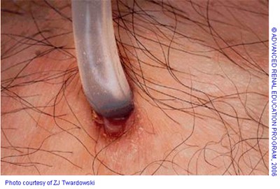
Grade 1: Good
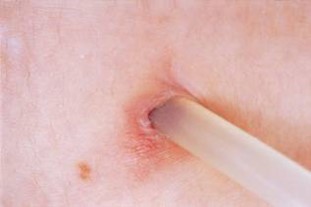
Photo courtesy of ZJ Twardowski, ©Advanced Renal Education Program, 2006
A good exit site usually takes more than 6 weeks of healing time to occur. The skin is natural in color and redness should not extend from the catheter more than 1-2 mm according to Teixidó’s guidelines. Twardowski suggests measuring the redness diameter from border to border, including the width of the catheter. Following the Twardowski guideline, 13 millimeters is the maximum diameter for redness measurement for an exit to be classified as “good.” There should be no pain, swelling, bright pink or red color, exuberant granulation tissue, external exudation, or abundant internal secretion.
Grade 2: Equivocal
An equivocal exit-site can be thought of as neither a good exit-site nor an obvious infection. In equivocal exit sites, purulent or bloody drainage is only present in the sinus and cannot be expressed outside and is accompanied by regression of the epithelium and slight exuberant granulation tissue in the sinus. There might be some mild redness extending two to three millimeters from the sinus edge to the border edge (according to Teixidó), but there is no pain, swelling or external drainage. Crust usually develops every one to two days. This crust may occur in the form of a cuff that is large or difficult to detach. Equivocal sites often suggest low-grade infections that may improve spontaneously or progress if left untreated.
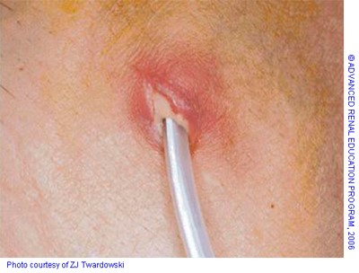
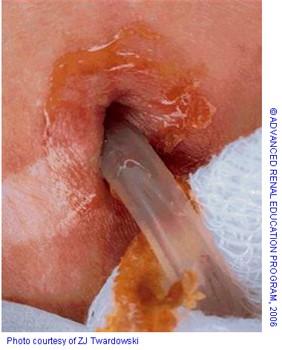
Acute exit-site infection
An acute infection is characterized by redness, swelling and tenderness. The measurement from the sinus edge to the border edge is greater than three to four millimeters. The outside diameter of the catheter is approximately five millimeters. Twardowski’s measurement suggests that the area of redness is greater than 13 mm in diameter. The erythema is more than twice the diameter of the catheter, and there is regression of the epithelium in the sinus. An acute infection is often painful and a scab might be present and/or daily crust. Scabs are composed of hardened serum and blood and can form as a result of capillary bleeding in the granulation tissue. Crusting alone does not mean infection. External drainage is purulent or bloody. This drainage may be spontaneous or may be expressed after pressing on the sinus. Purulent drainage may be present in the form of a white, yellow, or green liquid. In addition to these obvious characteristics, a large amount of serous drainage may also signal an infection. Purulent drainage should always be cultured. Although, positive cultures of normal-appearing exit sites indicate colonization but not infection.
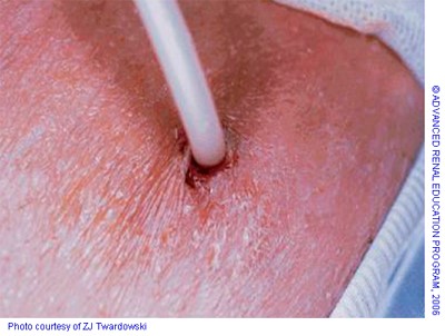
 Chronic exit-site infection
Granulation tissue is typically present both externally and in the sinus of the exit site in chronic infections. The exit is sometimes covered by a large, persistent crust or scab. There is usually no pain, redness or swelling, and the skin is often hyper-pigmented. Drainage from a chronically infected or Grade 4 exit site is the same as for the acutely infected or grade 3 exit site. It is important to note that the key distinction between the acute or Grade 3 and chronic or grade 4 infected sites is the duration of the infection.
 Tunnel infections
Tunnel infections are associated with redness, swelling and tenderness over the tunnel and may be accompanied by intermittent or chronic, purulent or bloody drainage that discharges spontaneously or after pressure on the cuff. These infections are often occult and are usually located between the internal and external cuffs. Ultrasonic evaluation of the tunnel is useful in confirming and assessing the extent of the peri-catheter abscess(5). Most, but not all, tunnel infections occur in conjunction with exit site infections. The presence of a tunnel infection increases the risk for peritonitis.
 A traumatized exit-site is not an infection but may involve pain, bleeding, scab development, and deterioration of the exit.
References
- Prowant BF, Twardowski ZJ. Recommendations for exit care. Perit Dial Int. 1996;16 Suppl 3:S94-S99. Available from: https://www.ncbi.nlm.nih.gov/pubmed/8860152.
- Twardowski ZJ, Prowant BF. Classification of normal and diseased exit sites. Perit Dial Int. 1996;16 Suppl 3:S32-S50. Available from: https://www.ncbi.nlm.nih.gov/pubmed/8860149.
- Teixidó J, Arias N, Tarrats L, Romero R. [The microbial pattern of the catheter exit-site infection in peritoneal dialysis: A non-diphtheria Corynebacteria emergence?]. Nefrol publicación Of la Soc Española Nefrol. 2007;27(3):350-358. Available from: https://www.ncbi.nlm.nih.gov/pubmed/17725455.
- Tiexidó J, Arias N. Catheter exit-site: photographic diagnostic table based on graded attributes (criteria) [Abstract]. Perit Dial Int J Int Soc Perit Dial. 1998;18(S1):S40.
- Buxo JAD, Black EB, Tyroler J. Ultrasonography in the diagnosis of peritoneal dialysis catheter tunnel abscess. Perit Dial Int. 1988;8(3):218. Available from: https://journals.sagepub.com/doi/pdf/10.1177/089686088800800310
P/N 102492-01 Rev A 07/2016