Hydrothorax
Hydrothorax is defined as the presence of excess fluid in the pleural cavity. The incidence of hydrothorax varies between 1.6-10% in PD patients, but predominantly in females. In addition, the right side appears to be more commonly affected than the left (1). A predisposing factor is polycystic kidney disease(2). Congenital causes include the localized absence of muscle fibers or tendinous diaphragmatic defects and eventration(1,3). Among the acquired etiologies are trauma to the abdomen and other causes of increased intra-abdominal pressure(4). A one-way passage of fluid between the peritoneal and pleural cavities is occasionally observed with valve-like defects of the diaphragm or hepatic capsule tamponade.
Diagnosis
Hydrothorax can be asymptomatic in about 25% of patients and is sometimes diagnosed as an incidental finding on routine physical examination(1,5). The most common signs and symptoms are dyspnea and inadequate ultrafiltration. Diagnosis confirmation may be obtained via a post infusion chest x-ray followed by a second simple chest x-ray after draining the peritoneal cavity. Isotope peritoneo-pleurograms with radioisotopes or peritoneography with contrast media can provide better definition of the diaphragmatic defect(1,6). In the absence of definitive evidence of a pleural-peritoneal communication, a pleural fluid sample can help to confirm the diagnosis by the presence of a higher glucose concentration than plasma (7), the LDH and protein concentrations of a transudate and the presence of d-isomers of lactate(3).
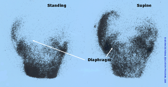
Figure 1: Pleuro-peritoneal leak

Diagnosis of a pleuro-peritoneal leak (hydrothorax) with scintigraphy is shown in figure 1. The left panel shows the patient in the standing position and minimal effusion above the diaphragm. The right panel shows the accumulation of fluid above the diaphragm when the patient assumed the supine position.
Treatment
The simple drainage of the peritoneal cavity and avoidance of overnight dwells in the supine position can correct the problem in some patients. Acute thoracentesis is seldom required for treatment of an acute and severe presentation of a hydrothorax with respiratory compromise. The persistence of hydrothorax requires permanent obliteration of the pleuro-peritoneal communication with pleurodesis using autologous blood, talc, tetracycline, surgical or thoracoscopic correction or video-assisted thoracoscopic (VATS) talc pleurodesis(1,3).
References
- Lew SQ. Hydrothorax: pleural effusion associated with peritoneal dialysis. Perit Dial Int. 2010;30(1):13-18. Available from: https://www.ncbi.nlm.nih.gov/pubmed/20056973.
- Fletcher S, Turney JH, Brownjohn AM. Increased incidence of hydrothorax complicating peritoneal dialysis in patients with adult polycystic kidney disease. Nephrol Dial Transplant. 1994;9(7):832-833. Available from: https://www.ncbi.nlm.nih.gov/pubmed/7970129.
- Tapawan K, Chen E, Selk N, Hong E, Virmani S, Balk R. A large pleural effusion in a patient receiving peritoneal dialysis. Semin Dial. 2011;24(5):560-563. Available from: https://www.ncbi.nlm.nih.gov/pubmed/21480997.
- Hausmann MJ, Basok A, Vorobiov M, Rogachev B. Traumatic pleural leak in peritoneal dialysis. Nephrol Dial Transplant. 2001;16(7):1526. Available from: https://www.ncbi.nlm.nih.gov/pubmed/11427671.
- Szeto CC, Chow KM. Pathogenesis and management of hydrothorax complicating peritoneal dialysis. Curr Opin Pulm Med. 2004;10(4):315-319. Available from: https://www.ncbi.nlm.nih.gov/pubmed/15220759.
- Bae EH, Kim CS, Choi JS, Kim SW. Pleural effusion in a peritoneal dialysis patient. Chonnam Med J. 2011;47(1):43-44. Available from: https://www.ncbi.nlm.nih.gov/pubmed/22111056.
- Davies SJ, Wilkie ME. Complications of Peritoneal Dialysis. In: Johnson RJ, Feehally JD, Floege J, eds. Comprehensive Clinical Nephrology. 5th ed. Philadelphia, PA: Saunders Elsevier; 2015:1107-1115.
The information and reference materials contained in this document are intended solely for the general education of the reader. It is intended to provide pertinent data to assist you in forming your own conclusions and making decisions. This document should not be considered an endorsement of the information provided nor is it intended for treatment purposes and is not a substitute for professional evaluation and diagnosis. Additionally, this information is not intended to advocate any indication, dosage or other claim that is not covered, if applicable, in the FDA-approved label.
P/N 102504-01 Rev. A 06/2016
