
Genital and Anterior Wall Edema
Genital and anterior wall edema are distressful complications for peritoneal dialysis patients. Despite its decreasing incidence over the years, approximately 10% of peritoneal dialysis patients may experience labial, scrotal and penile edema. According to evidence it appears that female patients have considerably lower incidence of this complication when compared to men. This unequal distribution can be the result of an embryonic remnant known as patent processus vaginalis(1). Patent processus vaginalis is present approximately in 80-95 % of all male newborns, however, its incidence decrease drastically in the first three years of age. Evidence shows that 20 % of those in whom the processus vaginalis remains patent will manifest signs and symptoms during their lifetime(2). Genital edema is commonly associated with anterior wall edema, the specific incidence of abdominal compromise is not exactly known, however, it is recognized to be less frequent than hernias in peritoneal dialysis patients(3).
Etiology
Clinicians have proposed two mechanisms related to pericatheter dialysate leakage and peritoneal tear(1,4).
- Dialysate can find its path through the soft-tissue plane from:
- The catheter insertion site
- Soft-tissue defect
- Peritoneal-fascial defect
- Dialysate can travel via a patent processus vaginalis to the scrotum or labia.
- In case viscera follows or accompanies the dialysate in its path through the processus vaginalis, an associated inguinal hernia will be present; in effect, when scrotal edema is diagnosed it is necessary to rule out the existence of an occult indirect inguinal hernia.
Risk Factors
Genital & Abdominal Edema: There is no general consensus on whether increasing the hydrostatic intraperitoneal pressure due to the presence of dialysis fluid in the peritoneal cavity increases the risk of abdominal wall complications. Some investigators found no more complications with higher pressures, while others observed increased hernia formation when the abdominal wall pressure was higher(5). However, Daugirdas et al. suggest that risk factors for genital and abdominal wall edema are similar to the ones for hernia formation, including(3):
- Large dialysate volume
- Sitting position
- Isometric exercise
- Valsalva maneuver
- Recent abdominal surgery
- Pericatheter leak & Hematoma
- Obesity
- Deconditioning
- Multiparity
- Congenital anatomical defects
Risk factors potentially related with the occurrence of dialysate leaks(6):
- Technique for peritoneal catheter placement
- Median surgical placement compared to paremedian implantation of peritoneoscopic insertion
- Initiation of peritoneal dialysis
- Immediate onset after catheter insertion vs. delay of 10-14 days
- Starting with higher dialysate volumes compared to 500ml increments
- Abdominal wall weakness
- Previous abdominal surgeries
- Multiple pregnancies
- Long-term therapy with steroids
- Hernias
- Heavy straining
- Obesity
Signs & Symptoms
Although genital edema is not harmful to the patient, it does cause considerable distress and discomfort. Typically, patients complain of rapid and persistent swelling of the scrotum and the penis. Discomfort, but minimal pain are referred, without urinary nor gastrointestinal symptoms. During physical examination, scrotal swelling and edema without redness or tenderness are commonly described (4). When the dialysate dissection compromised superficial planes of the abdomen, the abdominal skin can look pale and boggy, moreover, indentations made by clothing, or by the catheter lying across the abdomen appear more prominent and deeper(1).
Diagnosis
In occasions, patients may misinterpret edema occurrence as an indicative of general fluid overload and consequently they will try to ultrafilter more fluid. Patients who develop edema will look for medical attention complaining of reduced effluent return, which in this case is not a consequence of a high transporter state. Careful physical examination should be performed in patients presenting with genital edema or complaint of decreased effluent volumes. It is best to examine the patient in standing position to detect edema or asymmetry of abdomen. If abdominal edema is present, leakage is likely from the inguinal region proximities (1). Physical examination is not conclusive and its contribution depends in the extent of edema, therefore, in addition to physical examination, imaging techniques may be implemented to investigate and determine the source of leakage, however, recognition of the cause can be challenging.
Numerous diagnostic imaging techniques have been utilized with different degrees of success, including: peritoneal scintigraphy, ultrasonography, computed tomographic, and, most recently, magnetic resonance (4). Yet, the three more utilized imaging techniques are abdominal radiography, computed tomography (CT), and scintigraphy. Magnetic resonance imaging (MRI) technique can also detect the abnormalities within the peritoneal cavity (1,6). Among the most used techniques, computerized tomography with intraperitoneal instillation of dye in the dialysate appears to be the most convenient technique to demonstrate the source of leak, especially in patients with patent processus vaginalis or with occult hernia(1).
Figure 1. Patent processus vaginalis: Scintigram showing patent processus vaginalis (arrow).
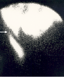
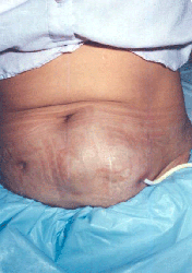
Figure 2. Pseudohernia: Image of asymmetrical pericatheter edema of the anterior abdominal wall due to pericatheter leak at the site of the internal cuff
(Picture used with permission)
Treatment
To treat genital edema, it is recommended to temporarily stop peritoneal dialysis. In addition, bed rest and scrotal elevation may help reduce the edema as well. If patient requires dialysis, frequent, low volume exchange on cycler or temporary hemodialysis can be utilized. Genital edema due to leakage through patent vaginalis can be treated with surgery. Finally, leakage from anterior abdominal wall may be managed by catheter replacement (1,7).
For abdominal edema, patient may need to interrupt peritoneal dialysis for 1-2 weeks or perform nocturnal dialysis with dry days. After edema is resolved, continuous ambulatory peritoneal dialysis (CAPD) may be reinitiated; however, some patient may require smaller volume therapy or cycler therapy in supine position to minimize intra-abdominal pressure (1,7).
Prevention
It is recommended that dialysis be initiated 10-14-days post-catheter insertion in order to avoid early dialysate leakage (8). If dialysis is required immediately, as in urgent start PD, the initiation in the supine position with reduced exchange volumes of 500-1500 mL has been recommended (6,9,10).
References:
- Bargman JM. Noninfectious Complications of Peritoneal Dialysis. In: Khanna R, Krediet RT, eds. Nolph and Gokal’s Textbook of Peritoneal Dialysis. Third. New York: Springer; 2009:571-609.
- Rahman N, Lakhoo K. Patent processus vaginalis: a window to the abdomen. Afr J Paediatr Surg. 2009;6(2):116-117. Available from: https://www.ncbi.nlm.nih.gov/pubmed/19661645.
- Blake PG, Daugirdas JT, Ing TS. Handbook of Dialysis. 4th ed. (Blake PG, Daugirdas JT, Ing TS, eds.). Philadelphia, PA: Lippincott Williams & Wilkins; 2007.
- Haggerty SP, Jorge JM. Laparoscopy to evaluate scrotal edema during peritoneal dialysis. JSLS. 2013;17(3):429-432. Available from: https://www.pubmedcentral.nih.gov/articlerender.fcgi?artid=3771763&tool=p….
- Del Peso G, Bajo MA, Costero O, Hevia C, Gil F, Díaz C, Aguilera A, Selgas R. Risk factors for abdominal wall complications in peritoneal dialysis patients. Perit Dial Int. 2003;23(3):249-254. Available from: https://www.ncbi.nlm.nih.gov/pubmed/12938825.
- Leblanc M, Ouimet D, Pichette V. Dialysate leaks in peritoneal dialysis. Semin Dial. 2001:50-54. Available from: https://www.ncbi.nlm.nih.gov/pubmed/11208040.
- Bargman JM. Hernias, Leaks, and Encapsulating Peritoneal Sclerosis. In: Daugirdas JT, Blake PG, Ing TS, eds. Handbook of Dialysis. fifth. Philadelphia: Wolters Kluwer; 2015:515-516.
- Crabtree JH. Peritoneal dialysis catheter implantation: avoiding problems and optimizing outcomes. Semin Dial. 2015;28(1):12-15. Available from: https://www.ncbi.nlm.nih.gov/pubmed/25338661.
- Povlsen J V, Ivarsen P. How to start the late referred ESRD patient urgently on chronic APD. Nephrol Dial Transplant. 2006;21 Suppl 2:ii56-ii59. Available from: https://www.ncbi.nlm.nih.gov/pubmed/16825263.
- Ghaffari A. Urgent-start peritoneal dialysis: a quality improvement report. Am J Kidney Dis. 2012;59(3):400-408. Available from: https://www.ncbi.nlm.nih.gov/pubmed/22019332.
The information and reference materials contained in this document are intended solely for the general education of the reader. It is intended to provide pertinent data to assist you in forming your own conclusions and making decisions. This document should not be considered an endorsement of the information provided nor is it intended for treatment purposes and is not a substitute for professional evaluation and diagnosis. Additionally, this information is not intended to advocate any indication, dosage or other claim that is not covered, if applicable, in the FDA-approved label.
P/N 102503-01 Rev A 06-2016